The NUT carcinoma remains a rare but highly aggressive malignancy and is often affecting younger patients, typically involving midline structures such as the head, neck and mediastinum. Although this carcinoma is uncommon in Australia and New Zealand, the importance of greater awareness and accurate identification strategies continue to grow as diagnostic capabilities advance across ANZ anatomical pathology laboratories.
To support pathologists in confidently and accurately detecting this entity, Cell Marque offers the NUTM1 (C52B1) rabbit monoclonal antibody which is designed to deliver strong nuclear staining performance with outstanding specificity.
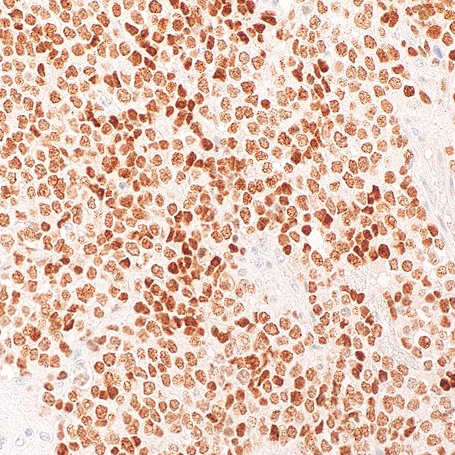
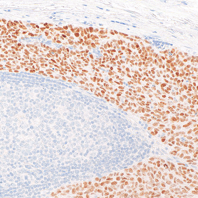
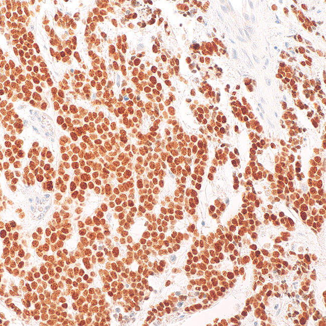
Cell Marque NUTM1 (C52B1) offers:
- Clear nuclear staining pattern for confident interpretation
- 87% sensitivity, and 100% specificity for NUT Carcinoma (Haack H, et al., 2009)
- A powerful adjunct to molecular confirmation where required
- Backed by Cell Marque quality, reliability and technical support
The NUTM1 antibody supports histopathology teams aiming to:
✔️ Improve recognition of NUT carcinoma in challenging cases
✔️ Strengthen diagnostic accuracy – NUT is a biomarker for NUT carcinoma and neoplasms harboring NUTM1 fusion genes; the presence of staining confirms the diagnosis and excludes potential mimics (Am J Surg Pathol 2009;33:984, Cell 2016;164:1060, Am J Surg Pathol 2018;42:636)
✔️ Support clinical teams with earlier and clearer answers – NUT is variably expressed among germ cell tumors, including dysgerminoma (64 – 93%), embryonal carcinoma (0 – 9%), immature teratoma (75%), seminoma (6 – 74%), spermatocytic seminoma (100%) and yolk sac tumor (7 – 10%); however, in this context and in the absence of a broader panel of immunohistochemical markers, NUT alone is of limited diagnostic value (Am J Surg Pathol 2009;33:984, Histopathology 2014;65:35, Pathology 2015;47:118)
THESE PRODUCTS ARE NOT AVAILABLE FOR PURCHASE BY THE GENERAL PUBLIC.