Making a difference to the people we help treat by creating value and improving healthcare outcomes through connecting innovative manufacturers with Australasian customers.
Autoimmunity
HEp-2 versus HEp-2000® – What is the difference?
What is the difference?
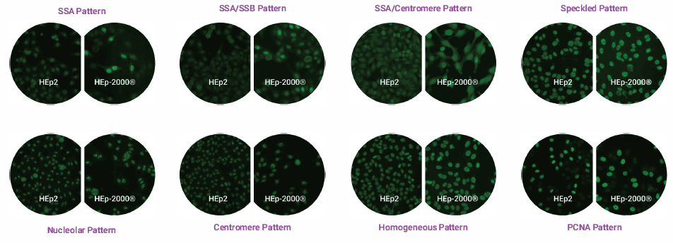
The HEp-2000® ANA Test System uses transfected mitotic human epithelioid cells (HEp-2). Historically, HEp-2 cells with mitotic figures have shown greater sensitivity and sharper pattern recognition than rodent kidney substrates. HEp-2000® contains HEp-2 cells transfected with multiple copies of the specific DNA sequence for the SSA/Ro autoantigen. Approximately 10-20% of the transfected cells over-express this antigen.
Detection of autoantibodies to SSA/Ro is more consistent with transfected HEp-2 cells compared to non-transfected ones. Autoantibodies to SSA/Ro may show a distinctive staining pattern on the transfected cells, which confirms the presence of anti-SSA/Ro antibodies. However, the absence of this distinctive pattern does not rule out the possible presence of these antibodies.
The key difference with HEp-2000® is its increased sensitivity in detecting and confirming antibodies to SSA/Ro compared to non-transfected HEp-2 cells. Numerous studies have consistently demonstrated the advantage of HEp-2000® over other methods for detecting anti-SSA/Ro antibodies. As long as slide-based assays remain the gold standard for ANA testing, HEp-2000® will continue to be the market leader in providing consistent and accurate clinical answers.
Reading and Reporting
All HEp-2000® cells are transfected to hyperexpress the 60kD SS-A/Ro antigen. However, only about 10% to 20% of the cells exhibit this hyperexpression. Consequently, when anti-SS-A/Ro antibodies are the only ANAs present, two distinct patterns may be observed:
Speckled Staining: All cells demonstrate speckled staining of the interphase cells. The 10% to 20% of cells hyperexpressing the antigen show stronger staining of the nucleus and/or nucleoli, with some cells potentially displaying cytoplasmic staining. This should be reported as ANA positive with two patterns: speckled and SS-A/Ro.
Distinctive SS-A/Ro Pattern: Only the hyperexpressing cells show staining of the nucleus, nucleoli, and possibly the cytoplasm, while the remaining interphase cells are negative. This should be reported as ANA positive with the SS-A/Ro pattern.
When the distinctive SS-A/Ro pattern is present, it confirms the presence of SS-A/Ro antibodies. In both cases, ENA testing is recommended to rule out the presence of autoantibodies against other ENAs.
The images below show the same sera testing on both HEp-2000® and HEp-2 slides.
Find out more about ANA HEp-2000® on the Immuno Concepts website