
Laboratories are dependent on using the tissue that they produce as the control for subsequent tests performed. Tissues, or more specifically the biomarkers of interest, are subject to preanalytical variations such as cold ischemic time and formalin fixation times.1 These, in combination with the actual processing of the tissue, are known to affect the quality of material which we have become more acutely aware of with digital pathology.2

With many biomarkers, particularly those that are used semi-quantitatively, obtaining a reliable or consistent expression is challenging. Human Epidermal growth Factor Receptor 2 (HER2) for example is often heterogeneously expressed. This in turn means that a sample selected based on the initial expression level may change rapidly. Image 1 shows relative heterogeneous expression of HER2 from a paper by Nitta et al Despite being amplified we see varied protein expression in close proximity.3
In combination, these factors mean that tissue can never be standardised. It is argued that using tissue fixed and processed in house as a control is deemed “best practise” (and previously laboratories were faced with no alternative), however, this is not entirely true. This is because when one considers that resected material is most frequently used as a control for biopsies or for referred cases, neither will have been fixed and processed identically to the control.
With clonal cell lines, one can standardise the growth, fixation and processing of the samples. Validated against commercially clinically validated assays and subsequently quality controlled with the same assays, enables the provision of samples with known levels of expression and reproducibility. In addition to analytical performance, with good cell morphology, one can determine appropriate localisation and other physical effects such as under/over retrieval. Image 2 shows controls placed on the top of the slide as an on-slide QC.

A closer look demonstrates appropriate staining for HER2 in image 3.
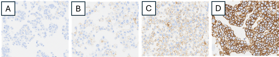
We can see appropriate 0, 1+, 2+ and 3+ expression in each of the cell lines. This consistent homogenous cell samples provides a level of confidence in assay performance not always entirely achieved with tissue (HER2 Analyte ControlDR, HCL028, HistoCyte Laboratories Ltd, Newcastle, UK). Furthermore, it allows a degree of trouble shooting previously hard to attain. In the new digital age, they lend themselves to measuring quality at a level never previously considered.
Read about The role of accurate diagnostics
Read about HistoCyte’s Cell line controls
View the HistoCyte Cell line controls Product Catalogue

The Issue with Tissue
Written by Colin Tristram
CEO, HistoCyte Laboratories Ltd
Director, The Immunohistochemistry Network
HistoCyte Cell line controls are for Research Use Only (RUO).
References:
- The Effect of Cold Ischemia Time and/or Formalin Fixation on Estrogen Receptor, Progesterone Receptor, and Human Epidermal Growth Factor Receptor-2 Results in Breast Carcinoma. Pekmezci M, Szpaderska A, Osipo C, Erşahin C.Patholog Res Int. 2012;2012:947041.
- Impact of Preanalytical Factors During Histology Processing on Section Suitability for Digital Image Analysis. Elizabeth A. Chlipala, Mark Butters, Miles Brous, Jessica S. Fortin, Roni Archuletta, Karen Copeland, Brad Bolon. Toxicol Pathol. 2021 Jun; 49(4):755-772.
- A gene-protein assay for human epidermal growth factor receptor 2 (HER2): brightfield tricolor visualization of HER2 protein, the HER2 gene, and chromosome 17 centromere (CEN17) in formalin-fixed, paraffin-embedded breast cancer tissue sections.
Request a Quote