
PRAME (Preferentially-expressed Antigen in Melanoma) is a gene encoded on the 22q11-22 chromosomal sequence, encoding a 509 amino acid residue protein.1
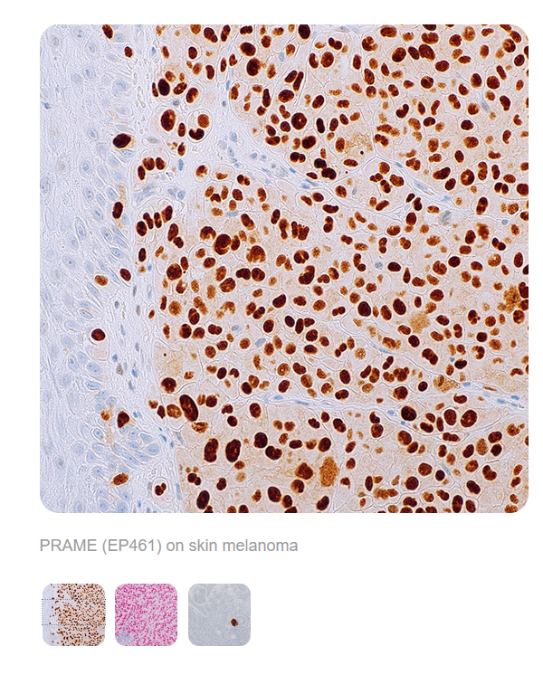
PRAME is a melanoma antigen that is preferentially expressed in tumours and is recognised by cytotoxic T lymphocytes.2,3 PRAME can be used to distinguish between malignant melanoma cells and nevus cells,4 and therefore may be useful for diagnostic purposes to support a suspected case of melanoma.
PRAME is considered a cancer-testis antigen (CTA)5 and is not strongly expressed in most other normal tissues. PRAME is positively expressed in about half of uveal melanomas,6 and the majority of mucosal melanomas7.
References
1. Wadelin, Frances et al. “Leucine-rich repeat protein PRAME: expression, potential functions and clinical implications for leukaemia.” Molecular cancer vol. 9 226. 27 Aug. 2010, doi:10.1186/1476-4598-9-226
2. Lezcano, Cecilia et al. “PRAME Expression in Melanocytic Tumors.” The American journal of surgical pathology vol. 42,11 (2018): 1456-1465. doi:10.1097/PAS.0000000000001134
3. Ikeda, H et al. “Characterization of an antigen that is recognized on a melanoma showing partial HLA loss by CTL expressing an NK inhibitory receptor.” Immunity vol. 6,2 (1997): 199-208. doi:10.1016/s1074-7613(00)80426-4
4. Lezcano, Cecilia et al. “Immunohistochemistry for PRAME in the Distinction of Nodal Nevi From Metastatic Melanoma.” The American journal of surgical pathology vol. 44,4 (2020): 503-508. doi:10.1097/PAS.0000000000001393
5. Zhang, Wa et al. “PRAME expression and promoter hypomethylation in epithelial ovarian cancer.” Oncotarget vol. 7,29 (2016): 45352-45369. doi:10.18632/oncotarget.9977
6. Gezgin, Gulcin et al. “PRAME as a Potential Target for Immunotherapy in Metastatic Uveal Melanoma.” JAMA ophthalmology vol. 135,6 (2017): 541-549. doi:10.1001/jamaophthalmol.2017.0729
7. Toyama, Aimi et al. “Analyses of molecular and histopathologic features and expression of PRAME by immunohistochemistry in mucosal melanomas.” Modern pathology : an official journal of the United States and Canadian Academy of Pathology, Inc vol. 32,12 (2019): 1727-1733. doi:10.1038/s41379-019-0335-4

THESE PRODUCTS ARE NOT AVAILABLE FOR PURCHASE BY THE GENERAL PUBLIC.